Reference: May 2024 | Issue 5 | Vol 10 | Page 40
Vitiligo is a chronic skin condition characterised by the loss of pigment-producing cells – called melanocytes – from the epidermis, leading to depigmented patches on the skin. The exact cause is unknown but is thought to involve a complex interplay of genetic, autoimmune, and environmental factors.1

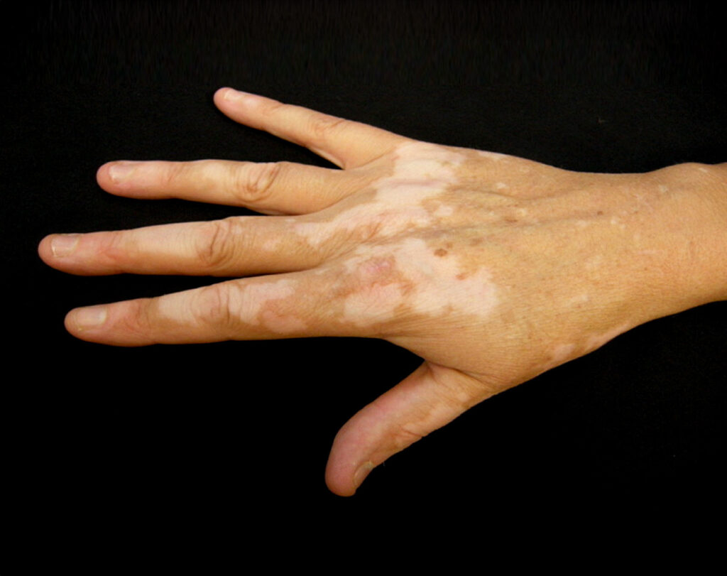
IMAGE 1: Vitiligo is a chronic skin condition that leads to depigmented patches on the skin
Its prevalence is approximately 0.1-to-2 per cent of the global population, including adults and children, and the disorder affects all races equally. Vitiligo can manifest at any age, although onset usually occurs in the second and third decade, before the age of 40. The condition can have a profound impact on quality-of-life due to its visible nature and potential psychological implications.2
Aetiology and pathogenesis
The aetiology of vitiligo is multifactorial and likely involves a combination of genetic predisposition, autoimmune mechanisms, oxidative stress, and environmental triggers. Genetic susceptibility is supported by familial clustering and genome-wide association studies.
Autoimmune mechanisms are implicated due to the presence of circulating autoantibodies, T-cell-mediated destruction of melanocytes, and the association with other autoimmune diseases such as thyroid disorders and alopecia areata.
Vitiligo often coexists with various autoimmune disorders, such as thyroid conditions like Hashimoto’s thyroiditis and Graves’ disease, as well as endocrinopathies like Addison’s disease and diabetes mellitus. Environmental factors such as stress, trauma, and certain chemicals may trigger or exacerbate vitiligo in genetically susceptible individuals.1,2,3
Clinical presentation
Clinically, vitiligo manifests as depigmented areas of skin with a convex border, without signs of inflammation, and is particularly noticeable in individuals with darker skin tones. The lesions are defined by clearly delineated, pearly white or depigmented areas in the form of macules and patches. They vary in shape, presenting as oval, round, or linear configurations, ranging from a few millimetres to centimetres in size. Over time, the patches tend to enlarge outward from their centre in a centrifugal manner.2,3
Vitiligo presents in various clinical forms, including trichrome, marginal inflammatory, and quadrichrome types. Koebner phenomenon is a commonly observed clinical feature, whereby vitiligo develops at sites prone to trauma such as cuts, burns, or abrasions. Initial lesions typically appear most commonly on the hands, forearms, feet, and face, with a tendency to have a periocular or perioral distribution.2,3
Vitiligo is categorised into three types based on its distribution and pattern: Generalised, segmental, and localised. Severity assessment is determined by the extent of affected body surface area. Progression of the condition is often unpredictable, and treatment outcomes vary among individuals.2,3
Complications of vitiligo
Ultraviolet (UV) exposure is one of the main risk factors for the development of skin cancer (nonmelanoma skin carcinoma). Because of a lack of melanin, affected skin is more vulnerable to the effects of sun exposure, and sunblock use on vitiligous areas is important to prevent sunburn/skin damage. Vitiligo may also be associated with eye problems such as inflammation of the iris, uveitis, and a partial loss of hearing (hypoacusis). Problems with confidence and self-esteem are common in people with vitiligo, particularly if it affects areas of skin that are frequently exposed.1,2,3
Diagnosis
The diagnosis of vitiligo relies primarily on clinical evaluation, with additional investigations carried out if necessary to confirm the diagnosis, rule out differential diagnoses, and assess for associated conditions. A comprehensive approach that considers both the physical and emotional aspects of the disease is important for effective management.
Clinical Examination: Doctors and dermatologists typically diagnose vitiligo based on the characteristic appearance of depigmented patches on the skin. Lesions are usually well-defined, with sharp borders and a loss of pigmentation. The distribution pattern of the patches may vary depending on the subtype of vitiligo (generalised, segmental, or localised).1,2,3
Wood’s lamp examination may be performed to enhance visualisation of depigmented areas. Under UV light, vitiligo lesions fluoresce bright white, aiding in the assessment of the extent of depigmentation.1,2,3
History and physical examination: A detailed medical history should be obtained, including the age of onset, progression of symptoms, family history of vitiligo, presence of autoimmune diseases, and exposure to potential triggering factors. Physical examination should assess the distribution of lesions, involvement of mucous membranes, presence of associated autoimmune conditions, and any evidence of Koebner phenomenon.1,2,3
Differential diagnosis: Hypo-pigmentary disorders, such as pityriasis alba, tinea versicolor, post-inflammatory hypopigmentation, idiopathic guttate hypomelanosis, drug-induced leukoderma, hypopigmented mycosis fungoides, scleroderma, and halo naevi should be considered and ruled out. Halo naevi are common through adolescence and have a striking clinical association with vitiligo.
Two main theories regarding the association between halo naevi and vitiligo exist: Halo naevi are a risk factor for developing vitiligo, and halo naevi are an early sign of vitiligo. However, some cases of vitiligo clearly spare melanocytic naevi, so the precise relationship between the two remains to be fully understood.
Mucosal vitiligo with isolated genital involvement should be evaluated with biopsy to rule out lichen sclerosus. Biopsy typically reveals the absence of melanocytes in the affected skin, and skin biopsy may be necessary in atypical cases to distinguish vitiligo from other conditions.4,5
Laboratory investigations: While there are no specific blood tests for diagnosing vitiligo, certain investigations may be warranted to evaluate for associated autoimmune conditions such as thyroid function tests and autoimmune antibody panels.
Screening for other autoimmune diseases commonly associated with vitiligo such as autoimmune thyroid disorders and pernicious anaemia, may also be carried out based on clinical suspicion. Thyroid-stimulating hormone, antithyroid peroxidase antibody, free T4, antinuclear antibody, full blood count with differential, and/or fasting blood glucose may be indicated to assess for comorbidities.1,2,3
Digital imaging and monitoring: Photography of vitiligo lesions at baseline and during follow-up visits can aid in monitoring disease progression and treatment response. Digital imaging techniques such as dermoscopy may provide additional insights into the morphology of vitiligo lesions and help differentiate them from other skin conditions.1,2,3
Psychological assessment: Given the visible nature of vitiligo and its potential impact on psychological wellbeing, a psychological assessment may be beneficial to evaluate the patient’s emotional state, body image concerns, and quality-of-life. This is particularly important in managing the psychosocial aspects of the disease.6,7,8
Treatment and management
Treatment of vitiligo aims to halt disease progression, induce re-pigmentation, and improve cosmetic appearance. Various treatment modalities are available, and selection is based on factors such as disease extent, location, and patient preference. Topical, systemic treatment, and phototherapy are useful for stabilisation and re-pigmentation of vitiligo.
For rapidly progressive disease, low-dose oral glucocorticoids, and phototherapy can be useful. Therapeutic options for stable, segmental vitiligo include topical therapies (eg, topical corticosteroids, topical calcineurin inhibitors), targeted phototherapy, and surgical therapy (tissue grafts and cellular grafts).
In recent years, efforts have been made to improve the re-pigmentation of vitiligo phototherapy by combination therapies, including narrowband (NB)-UVB with glucocorticoids and topical calcineurin inhibitors. Targeted therapies such as biologics targeting cytokines and small-molecule inhibitors targeting intracellular signalling molecules, are recently emerging as promising therapeutics for autoimmune diseases.9,10
Topical therapies
Corticosteroids: Topical corticosteroids (TCS) are commonly used for localised vitiligo. They work by suppressing inflammation and the immune response in the affected skin, thus promoting re-pigmentation. TCS, either potent (betamethasone valerate) or very potent (clobetasol propionate), are considered first-line therapy for vitiligo.9
Calcineurin inhibitors: Tacrolimus and pimecrolimus are topical calcineurin inhibitors that can be used as alternatives to corticosteroids, particularly in sensitive areas such as the face and genitals. They modulate immune responses and help promote re-pigmentation.9
Vitamin D analogues: Calcipotriol, a synthetic vitamin D analogue, may be used alone or in combination with other topical agents to enhance re-pigmentation. A greater efficacy rate is achieved when using combination therapy methods rather than relying on one form of treatment.9
Phototherapy
NB-UVB phototherapy: Narrowband (NB)-UVB is considered the gold standard phototherapy for vitiligo. It involves exposing the affected skin to UVB light of specific wavelengths, which stimulates melanocyte proliferation and migration, leading to re-pigmentation.2,3,9
Psoralen with UVA (PUVA) therapy: PUVA involves combining oral or topical psoralen with UVA exposure. Psoralen sensitises the skin to UVA radiation, enhancing re-pigmentation. PUVA is often reserved for cases resistant to NB-UVB or when vitiligo is widespread.9
Excimer laser therapy
Excimer lasers deliver targeted UVB radiation to depigmented areas, allowing for precise treatment with minimal exposure to surrounding healthy skin. Excimer laser therapy is painless, effective for localised vitiligo, and can be used in combination with other treatments.9
Surgical interventions
Surgery is only an option for vitiligo that is segmental or stable. Skin grafting and micropigmentation are the most common surgical procedures.
Autologous melanocyte transplantation: In this procedure, melanocytes are harvested from a donor site, such as the patient’s unaffected skin, and transplanted to depigmented areas. This technique is effective for stable, localised vitiligo.2,3,9
Punch grafting: Punch grafting involves transplanting small pieces of skin containing melanocytes into depigmented areas. It is suitable for stable vitiligo with well-defined lesions.3
Combination therapies
Combining different treatment modalities, such as topical therapies with phototherapy or surgical interventions, may enhance treatment efficacy, particularly in refractory cases.2,3,9 Emerging therapies for the condition include:
Janus kinase (JAK) inhibitors: JAK inhibitors, such as tofacitinib and ruxolitinib, are being investigated for their potential in treating vitiligo. These medications target inflammatory pathways involved in autoimmune diseases.9,10
Biologic agents: Biologic drugs targeting specific immune pathways, such as anti-tumour necrosis factor agents (eg, adalimumab) or anti-interleukin-17 agents (eg, secukinumab), show promise in early clinical trials for treating vitiligo.9,10
Emerging therapeutics targeting microRNAs (miRNAs): miRNAs may be involved in vitiligo pathogenesis via the modulation of vital gene expression in melanocytes and serve as novel therapeutic targets for vitiligo therapy.10
Emerging therapeutics targeting regulatory T-cells (Tregs): Tregs are a suppressive CD4+ T-cell subset that possesses a capacity to suppress self-reactive T-cell activation and expansion. A clear decrease in Treg cells has been observed in vitiligo skin within lesional, non-lesional, and perilesional sections, indicating that increasing the number of Tregs with normal function might be an important therapeutic intervention for vitiligo treatment.10
Supportive therapies
Camouflage: Cosmetic and camouflage techniques, which can be permanent (tattoos) or temporary (liquid dyes, traditional remedies, foundation-based cosmetic camouflage, and self-tanning products), can help conceal depigmented areas, improving the cosmetic appearance and quality-of-life for individuals with vitiligo.6,7
Psychological support: Counselling and support groups can assist patients in coping with the emotional impact of vitiligo and managing body image concerns. The social and psychological ramifications of the condition can be profound, and patients have reported associated feelings of embarrassment, anxiety, shame, and depression. In some cultures, vitiligo is a highly stigmatised disorder.8
The treatment of vitiligo is multifaceted, with various therapeutic options available depending on the extent and severity of the disease. Advances in research continue to expand treatment options, offering hope for improved outcomes and quality-of-life for individuals affected. Treatment plans should be individualised based on patient characteristics and preferences, with regular follow-up to monitor response and adjust therapy as needed.9
Treating vitiligo can be challenging as it often involves inconsistent clinical outcomes and a tendency for the condition to relapse. Therapy should be tailored to the individual, considering factors such as the type of vitiligo, its activity, and the potential side-effects of the chosen treatment.
It is important to note that all available therapies for vitiligo have their limitations, and there is currently no treatment known to consistently achieve re-pigmentation in all patients.
Prognosis
Vitiligo typically shows an insidious onset, and the natural progression of the disease is unpredictable. It may show slow spread with periods of stabilisation or rapid evolution. Vitiligo may stabilise for years only to progress without clear cause. The prognosis depends upon the age of onset and the extent of disease.
Early disease onset is usually associated with the involvement of greater body surface area and rate of progression. Few types and certain locations may be responsive to treatment. Refractory cases have been noted in patients presenting with segmental vitiligo and younger than 14 years of age. Most patients on treatment usually experience intermittent cycles of pigment loss and disease stabilisation.
Despite advancements in understanding its underlying causes, there remains a need for more effective and targeted treatment options. Continued research efforts aimed at uncovering the underlying mechanisms and developing novel therapies are important to improve outcomes and quality-of-life for patients with vitiligo.6,9,11
References
- Ahmed jan N, Masood S. Vitiligo. Florida: StatPearls Publishing; 2024. Available at:
www.ncbi.nlm.nih.gov/books/NBK559149/. - Bergqvist C, Ezzedine K. Vitiligo: A review. Dermatology. 2020;236(6): 571-592.
- Joge RR, Kathane PU, Joshi SH. Vitiligo: A narrative review. Cureus. 2022;14(9): e29307.
- Dina Y, McKesey J, Pandya AG. Disorders of hypopigmentation. J Drugs Dermatol. 2019;18(3): s115-s116.
- Cohen BE, Manga P, Lin K, et al. Vitiligo and melanoma-associated vitiligo: Understanding their similarities and differences. Am J Clin Dermatol. 2020;21(5): 669-680.
- AL-smadi K, Imran M, Leite-Silva V, et al. Vitiligo: A review of aetiology, pathogenesis, treatment, and psychosocial impact. Cosmetics. 2023;(10): 84.
- Grimes PE, Miller MM. Vitiligo: Patient stories, self-esteem, and the psychological burden of disease. Int J Womens Dermatol. 2018;4(1): 32-37.
- Bibeau K, Ezzedine K, Harris JE, et al. Mental health and psychosocial quality-of-life burden among patients with vitiligo: Findings from the Global VALIANT Study. JAMA Dermatol. 2023;159(10): 1124-1128.
- Kubelis-López DE, Zapata-Salazar NA,
Said-Fernández SL, et al. Updates and new medical treatments for vitiligo (Review). Exp
Ther Med. 2021;22(2): 797. - Feng Y, Lu Y. Advances in vitiligo: Update on therapeutic targets. Front Immunol. 2022;13: 986918.
- Stiegler J, Brickley S. Vitiligo. A comprehensive overview. J of the Derm
Nurses’ Ass. 2021;13(1): 18-27. - Dermnet New Zealand. Hand vitiligo image. Florida: StatPearls Publishing; 2024. Available at: www.ncbi.nlm.nih.gov/books/NBK559149/figure/article-31232.image.f1/?report=objectonly.
