


Phlebotomy is a crucial diagnostic and therapeutic tool in clinical practice. The indications for phlebotomy are numerous, and countless medical conditions can be diagnosed and treated based on the results of a patient’s blood tests. Diagnostic phlebotomy is used constantly in clinical environments to increase the understanding of a patient’s clinical presentation and improve their clinical outcomes.
Therapeutic phlebotomy is a treatment procedure performed for certain blood disorders such as haemochromatosis and polycythaemia vera, in which the removal of red blood cells or serum iron is the most efficient method for managing the condition’s symptoms and complications.
Education and training
Venepuncture is one of the most commonly performed invasive procedures, and is routinely undertaken by nurses, doctors, and phlebotomists. All healthcare professionals undertaking phlebotomy must be trained and competent in venepuncture, and the HSE has produced a Guiding Framework for Education, Training, and Competence Validation in Venepuncture and Peripheral Intravenous Cannulation for Nurses and Midwives. The HSE venepuncture programme or eLearning module consists of five units containing learning outcomes, theoretical content, self-assessment questions, and resources provided for supplementary reading.
The theoretical and self-assessment component of the programme is completed through www.hseland.ie. The clinical knowledge assessment is a multiple-choice examination of one-hour duration. The supervised clinical demonstration and practice session is facilitated through the Centres of Nursing/Midwifery/Children’s Nurse Education, practice development units, or designated educational providers, and is recommended as a four-hour session. Supervised clinical practice assessments, and the final competence assessment, must be undertaken in the clinical area and be completed within a 12-week timeframe from commencement of the eLearning module/theoretical component.
A certification of competence is issued by the educational provider on receipt of the final competence assessment form and skill completion checklist. The National Standardised Certificate of Competence Achievement enables transferability and recognition of venepuncture skills across the health service. Upon completion of the skill pathway, each nurse or midwife should have the knowledge, skills, and competence to practise the skill of venepuncture across healthcare organisations. In order to maintain competence, it is recommended that up to 10 venepuncture procedures per month is required or as predefined in local organisational policy. It is also important to keep up-to-date with any changes in best practice, local policy, and equipment utilised for the procedure.
In order to perform venepuncture safely, the practitioner must have knowledge of: The relevant anatomy and physiology; the criteria for choosing both the vein and devices to use; potential problems which may be encountered; how to prevent them and necessary interventions; the health and safety risk management of the procedure; and the correct disposal of equipment. Adherence to aseptic techniques must be applied. It is the responsibility of the healthcare practitioner carrying out phlebotomy to ensure that the procedure is carried out correctly and competently according to the phlebotomy clinical guidelines, and in accordance with their professional accountability and scope of practice.
Preparation and assessment
Patient consent and cooperation are important factors in any clinical consultation and the patient should be provided with clear information about the venepuncture procedure and the blood tests being carried out. At the outset of the phlebotomy consultation, it should be established whether the patient has had blood taken before, has any allergies and/or needle phobias, or has ever fainted during previous injections or blood draws. If the patient is anxious or afraid, it is important to provide reassurance and make them comfortable.If the patient has a history of fainting during venepuncture, they should lie down for the procedure. Good lighting and positioning of the patient is important to assess the veins and carry out the phlebotomy procedure.
A patient assessment provides the information necessary to assess the size, position, and condition of the patient’s veins, and make the appropriate equipment choices. It is important to inspect and palpate the patient’s veins to assess if they are visible, close to the surface or deeper, small and fragile, or large and supple. Some patients may have hardened or sclerotic veins, and these should be avoided because they are difficult to puncture and any attempt can lead to further patient harm. Injury, disease or treatment, eg amputation, fracture, stroke, or mastectomy, and axillary node dissection may prevent the use of a certain limb for venepuncture.
The age and weight of a patient will also influence the choice of vein. Care must be taken with fragile veins, and the largest vein should be chosen along with the smallest gauge device to reduce the amount of trauma to the vessel. Other factors that can cause poor superficial venous access are dehydration and shock, while the temperature of the environment can influence venous dilation. Venepuncture itself may cause the vein to collapse or go into spasm, producing discomfort and a reduction in blood flow. Careful preparation and choice of vein will reduce the likelihood of this happening, and stroking the vein or applying heat can help resolve it.
Most blood draws are performed in the antecubital space of the patient’s arm. If there is not a suitable vein in the antecubital space, the back of the patient’s hand is also acceptable. Tourniquets are a potential source of infection, with up to 25 per cent of tourniquets contaminated through lack of hand hygiene on the part of the clinician or reuse of tourniquets. To avoid contamination, tourniquets should be single use, visibly clean before use on the patient, and should never be reused.
Once the tourniquet is in place, the patient should be asked to make a fist and hold it. The patient should not be asked to pump his or her fist because this may cause a change in the blood composition and lead to erroneous results. The median cubital vein is the first choice for drawing blood because it has a decreased proximity to arteries and nerves in the arm. The more lateral cephalic vein is the second choice, and the basilic vein in the medial arm is the last choice. The basilic vein lies over a major artery and a nerve in the arm, which increases the risk of patient injury.
For veins that are small, difficult, and close to the surface, such as those on the back of the hand, the best choice is a butterfly or winged needle set. A butterfly needle is shorter, with a smaller diameter (23-to-25 gauge), and is easily controlled. Butterfly needles also have a length of small, clear tubing between the needle that accesses the vein and the needle that attaches to the tube holder. When the needle is inserted into the vein, a blood flashback will be visible in the tubing, making it easier to recognise that the vein has been accessed.
Blood collection bottles
Blood tests are routinely collected to define baseline results, confirm disease, monitor disease, and regulate therapy treatment. It is important that the correct blood tube is used for each test. Blood tests are sent to various departments within the laboratory such as haematology, biochemistry, and microbiology.Using the correct type of blood sample tube, filling it to the right level, and in the correct order are important for accurate test results. Tube types are indicated by colours that are universally associated with a certain additive or tube property, depending on whether the blood is being collected for a plasma or a serum sample.
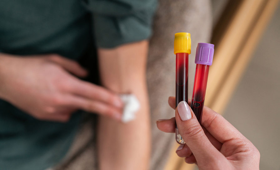
Vacutainer blood bottles and cap colours
Yellow: Clotted anticoagulant(no anticoagulant). Test: All serum tests.
Grey: Anticoagulant: Fluoride oxalate. Test: Blood glucose.
Purple: Anticoagulant:Ethylenediaminetetraacetic acid (EDTA). Test: Full blood cell count (FBC)/erythrocyte sedimentation rate (ESR).
Green: Anticoagulant:Lithium heparin. Test: Plasma tests.
Blue: Anticoagulant:Sodium citrate. Test: Coagulation tests.
Pink: Anticoagulant: EDTA. Test: Cross match.
Plasma contains clotting proteins and serum does not. If a test requires a whole blood or plasma sample, eg, FBC, then the tube needs to contain an anticoagulant additive to prevent clotting. When obtaining a serum sample, eg, serum electrolytes, the blood needs to clot within the tube before centrifugation and serum collection. In this case, the tube has no additive or contains a clot activator. Centrifugation is the process in the lab where the sample tubes are placed in a machine that spins at a high number of revolutions per minute. The spinning action causes the heavier formed elements such as blood cells and platelets to consolidate at the bottom of the tube. The liquid portion of the sample, serum or plasma, is less dense, and therefore pools in the upper part of the tube, making it easier to collect. Whether the sample is plasma or serum, there are multiple tube types that also have separator gels present in the bottom of the tube. The separator gel migrates to a position between the formed elements, and the plasma or serum, during centrifugation. The gel creates a barrier that helps prevent whole cells from contaminating the serum or plasma sample.
The use of vacuum extraction tube systems as closed systems for blood collecting reduces the risk of direct exposure to blood and has made it easier to take multiple samples from a single venepuncture. Sample tubes should fill to the appropriate amount automatically because of the vacuum in each tube. It is important not to remove the tube before it is full because it can affect the additive-to-blood ratio in the tube. When switching tubes, stabilise the tubeholder with the non-dominant hand, making sure not to change the needle insertion depth. When removing the filled tube and placing the new tube, use the two flanges on the side of the tubeholder to stabilise it and push the new tube onto the needle. Tubes are filled in a specific order to avoid additive contamination from tube to tube. Depending on the lab tests being collected, as well as facility protocol, a waste or discard sample may be required as the first tube.
If a blood sample is poorly collected, the results may be inaccurate and misleading, and the patient may have to undergo the inconvenience of repeat testing. Three major issues resulting from errors in blood collection are haemolysis, contamination, and inaccurate labelling.
Adult peripheral venepuncture guidelines
Equipment:
– Clinical tray with disposal sharps unit;
– Non-sterile powder-free disposable gloves;
– Disposable tourniquet (single use);
– Large 70 per cent isopropyl alcohol swabs;
– Gauze swabs;
– Sterile disposable double-ended vacutainer needle/plastic vacutainer holder and vacuette winged blood-collecting device;
– Appropriate blood specimen tubes;
– Sterile adhesive plaster or hypoallergenic tape;
– Laboratory specimen requisition forms;
– Plastic specimen laboratory bag or biohazard bag (where applicable).
Procedure:
1. Explain the procedure to the patient and gain informed consent.
2. Wash hands thoroughly using correct hand hygiene techniques and dry with paper towel.
3. Apply the tourniquet. The tourniquet should be tight enough to impede venous return while not affecting arterial flow. Allow time for the veins to fill. Ensure that the tourniquet is not applied for longer than clinically indicated.
4. Make a full assessment of the patient’s veins. Inspect the area for phlebitis, infection or oedema, bruised or inflamed veins, or any veins that have undergone multiple punctures. These veins are to be avoided.
5. Palpate the veins most likely to be used. Stroking the vein and observing the venous refills are helpful in determining the condition of the vein.
6. Assess and select the most appropriate vein.
7. Select the device based on vein size and location.
8. Decontaminate hands using correct hand hygiene techniques.
9. Put on well-fitting powder-free disposable gloves.
10. Clean the insertion site using a 70 per cent isopropyl alcohol-impregnated swab for a minimum of 30 seconds and allow to air dry for a minimum of 30 seconds.
11. Do not re-palpate or touch the skin.
12. Remove the needle from the packaging and inspect it for any faults.
13. Stabilise the vein by applying manual traction on the skin a few centimetres below the proposed insertion site using the non-dominant hand.
14. Ensure the needle tip is in the bevel-up position. Place the device directly over or to the side of the vein and insert the needle smoothly through the skin at a 30-degree angle, according to the depth of the vein.
15. As soon as puncture of the vein wall is felt, level off the needle by decreasing the angle of the needle to the skin. If using a winged blood collection device, a flashback of blood is seen in the tubing.
16. Slightly advance the needle in order to stabilise it within the vein if possible.
17. Gently release the skin tension. If using a winged device, tape one wing to stabilise the device if necessary.
18. Do not exert any pressure on the needle.
19. Keeping the needle and vacutainer holder steady in position, attach the appropriate blood specimen tube onto the needle inside the holder and withdraw the blood.
20. Samples may be obtained one after another by removing the filled tube and replacing it with another. Ensure that the tube is correctly filled to the fill mark on the tube label.
21. Collect samples in the following order, as recommended by manufacturer’s guidelines:
1 Non-additive tube;
2 Coagulation;
3 Serum;
4 Heparin;
5 EDTA;
6 Glucose;
7 All others.
22. During filling of the last tube, release and remove the tourniquet to decrease the pressure within the vein.
23. It may be necessary to release the tourniquet at the beginning of the sampling as inaccurate measurements may be caused by haemostasis. This is specific when drawing blood to assess calcium levels.
24. Remove the last specimen tube from the holder, prior to removing the needle.
25. Place a gauze swab over the puncture point. Remove the needle but do not apply pressure until the needle has been fully removed.
26. Apply firm digital pressure directly over the puncture site until haemostasis is achieved.
27. The patient may apply pressure with the fingers but should be discouraged from bending the arm if a vein in the antecubital fossa is used.
28. After a sample has been taken, it is important that the sample be gently inverted as per vacutainer manufacturer mixing guidelines (Figure 2). This will allow the sample to mix with the additive and reduce the risk of clots forming in the tube, which can later break during centrifugation and cause haemolysis.
29. All specimen tubes must be labelled immediately with the relevant patient details.
30. Inspect the puncture site before applying a dressing.
31. Apply an adhesive plaster or dressing.
32. Ensure the patient is comfortable and pain free.
33. Discard waste, making sure it is disposed of in the correct sharps-containers and appropriate waste bags.
34. Remove gloves and dispose of appropriately. Wash and dry hands thoroughly.
35. Place specimens in a sealable section of the plastic laboratory transport bag. Place completed blood request forms in the appropriate pocket of laboratory transport bag.
36. Follow local procedures for collection and transport of specimens to the laboratory.
Complications
Complications that may occur when venepuncture is performed include arterial puncture, nerve injury, haematoma, syncope, and infection. The most common adverse event for the patient is the development of a haematoma at the blood drawing site.In most cases, haematomas are mild, although severe cases may occur involving damage to the surrounding tissues, nerves, and blood vessels. The risk of a haematoma can be reduced by applying pressure to the drawing site, and pressure and an ice pack should be used to slow the bleeding if a haematoma occurs. Another common adverse effect is syncope. If the patient faints during phlebotomy, the tourniquet should be immediately released, the needle should be removed (or in therapeutic phlebotomy procedures the catheter should be capped), and appropriate treatment used to manage the symptoms.
Another adverse risk of phlebotomy is needle-stick injuries among healthcare workers and these injuries are associated with a risk of HIV, hepatitis B, hepatitis C, and other pathogen transmissions. Clinicians and phlebotomists should be well acquainted with the possibility of needle-stick injuries and blood-borne pathogen exposure, and take appropriate measures to avoid these injuries and exposures. Regulations and guidelines for phlebotomy should include precautions regarding needle-stick injuries, the immediate treatment steps, post-exposure prophylaxis/serological testing to exclude infection, vaccination or medications, and a notification system to alert the appropriate infection control centre.
Therapeutic phlebotomy
Therapeutic phlebotomy is beneficial for certain conditions, and is the preferred treatment for blood disorders in which the removal of red blood cells or serum iron is the most efficient method for managing the symptoms and complications. Therapeutic phlebotomy is indicated for the treatment of hemochromatosis, polycythaemia vera, porphyria cutanea tarda, sickle cell disease, and non-alcoholic fatty liver disease with hyperferritinaemia.
In Ireland, patients undergo therapeutic phlebotomy for haemochromatosis in a variety of settings, including acute clinics in hospitals, the Irish Blood Transfusion Service, general practice, and GP minor surgery networks. Ireland has the highest reported prevalence of hereditary haemochromatosis (HH) in the world. The treatment of HH consists of life-long phlebotomy therapy and monitoring of iron indices, in particular serum ferritin. The advocated standard practice is to maintain serum ferritin at 50-100µg/L.
In January 2016, a survey sent to the Hospital Groups highlighted that the majority of patients were being treated in the acute sector, typically within a day-ward setting, with a smaller number seen in outpatient clinics. Haemochromatosis services have traditionally been run through endoscopy units, diabetic day centres, IV therapy suites, gastroenterology and hepatology clinics, and haematology day wards, with patients referred by their GP or from another hospital service for both diagnostic work-up and therapeutic management. Most services are consultant-led, nurse-delivered, ambulatory services. In general, staff nurses are accountable and responsible for the day-to-day management and operation of the phlebotomy service, with support and guidance from consultant gastroenterologists and/or hepatologists.
In 2019, terms of agreement between the Department of Health, the HSE, and the Irish Medical Organisation regarding GP contractual reform and service development decided that therapeutic phlebotomy for HH be added to the general medical services (GMS), and that current admissions of GMS/GP visit card (GPVC) patients to the acute setting and outpatient department be treated in general practice.
According to the agreement:“The defined target population of patients is predominately aimed at men, aged between 40 and 65 years. The prevalence rate of haemochromatosis in Ireland is estimated at 55,000. Of this figure, approximately 20,000 are women who do not require venesection, and another 15,000 males who, while having the condition, will not require venesection.
“In addition, it is estimated there are approximately 5,000 patients whose condition may remain undiagnosed. That leaves approximately 15,000 patients requiring venesection, of which it is estimated between 7,000 and 8,000 will be GMS/GPVC patients with an average of three visits per patient per year.”
Some GP practices currently do provide therapeutic phlebotomy services for HH, however, many still do not. While time consuming, treatment for HH is feasible and general practice is particularly amenable to diagnosis, treatment, and ongoing follow-up in the primary care setting. Advantages are that patients can be managed in the community out of the acute setting, close to the patient’s home with the GP in charge of both the clinical management and treatment of the patient.
Therapeutic phlebotomy for patients with HH involves the removal of 400-500ml of blood (200-250mg iron). The procedure is similar to that of blood donation. The objectives are:
– Fasting serum ferritin (SF) 50-100µg/L;
– Fasting transferrin saturation (TS);
– Avoid anaemia;
– Allow haematocrit/haemoglobin to fall by no more than 20 per cent of prior level.
Patients are eligible for therapeutic phlebotomy for HH if they have the HFE gene and increased body iron stores (fasting SF >200µg/L in pre-menopausal women, and >300µg/L in men and post-menopausal women, and fasting TS ≥45 per cent).
The patient should also have had an FBC in the past three months; liver function test (serum AST and ALT activity); fasting blood glucose level or haemoglobin A1C (HbA1c); and documentation of any specific risks such as HIV, hepatitis, and methicillin-resistant Staphylococcus aureus (MRSA).
Therapeutic phlebotomy treatment regimens are individualised to each patient, and patient factors such as age, gender, weight, general health, and likelihood of compliance need to be considered.
Special precautions may also need to be taken for patients on beta-blockers as they may be susceptible to syncope. It requires around 30 minutes to undertake therapeutic phlebotomy.
Therapeutic phlebotomy procedure prerequisites
– Correct patient (check name and date of birth);
– Confirmed diagnosis of HH;
– Stable haemoglobin >12.5g/dL;
– Serum ferritin above 25μg/L;
– Stable blood pressure systolic 110-160mmHg, diastolic 60-95mmHg;
– Stable pulse 50-100/minute;
– Ability to appropriately dispose of collected blood (clinical waste/biohazard);
– Venous access – usually cubital fossa of the opposite side from the most recent phlebotomy;
– Adequate pre-phlebotomy hydration;
– Check with the patient regarding recent oral intake, it is best if food is eaten two hours before the procedure;
– Explain procedure to the patient and ensure the patient understands it;
– Consent obtained (both verbal and written).
Possible complications during therapeutic phlebotomy include haematoma, hypovolaemia, vasovagal syncope, venous scarring, phlebitis, and adverse reaction to lignocaine if used. The patient must be observed at all times, and if they become tachycardic, hypotensive, restless or clammy, the procedure must be stopped and the patient reviewed.
It is rare for patients not to tolerate therapeutic phlebotomy, except for some with severe cardiac disease, anaemia, or hypoproteinaemia. l
References on request


Leave a Reply
You must be logged in to post a comment.