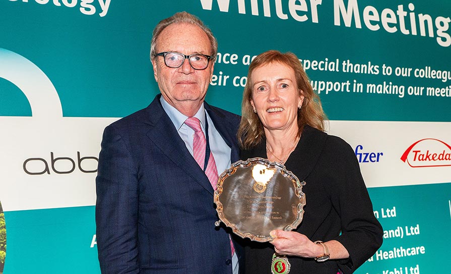
Irish Society for Rheumatology Spring Meeting, Virtual, 13 May 2021

Prof Patrick Kiely, Consultant Rheumatologist at St George’s University Hospital, London, UK, is leading a European League Against Rheumatism (EULAR) initiative to establish guidelines for haemochromatosis arthropathy in association with the Mater Misericordiae University Hospital, Dublin, and the ISR. As such, it was fitting that he presented at the ISR Spring Meeting on the topic.
Prof Kiely shared how he established a clinic for haemochromatosis arthropathy at St George’s in 2012, with the aim of finding out more about the condition, which currently takes between seven-to-eight years from first presentation to diagnosis. To date, he has seen 135 patients from across the country and was able to gather data from these patients. In association with the UK Haemochromatosis Society, Prof Kiely gathered 470 responses to a widespread patient survey, the majority of whom were UK-based. The results of this study revealed that joint pain and fatigue were, by far, the most prevalent symptoms present at the time of diagnosis. The areas most affected by the condition are the knee, ankle, metacarpophalangeal joint, and proximal interphalangeal joint in descending order.

Examining the ankle in more detail, ankle osteoarthritis (OA) is rare and while Prof Kiely’s research revealed that 35 per cent of patients self-reported symptoms in the ankle, a cross-sectional study of 199 patients in Germany and Austria had a similar finding of 32.9 per cent. On further examination Prof Kiely noted that research out of the US found that only 7.5 per cent of all cases of Kellgren grade 3 or 4 OA in a particular centre in the US in one year were ankle-related. The majority of hip and knee OAs were primary, but the ankle cases were post-traumatic. Follow-up data collected over the next 13 years revealed the same findings, the majority of OA ankle cases are post-traumatic.
Following on from this, and Prof Kiely’s repeated observation regarding ankle cysts that resembled “bunches of grapes”, he embarked on a research topic, which asked: “Are there features on MRI that could distinguish HA [haemochromatosis arthropathy] from primary OA in the hindfoot?” The study comprising 30 hindfoot MRIs from 22 genetic haemochromatosis patients found that this group have a higher score at the ankle and middle facet subtalar joints for OA features, including large bone marrow lesions or cysts, extensive cartilage loss and large osteophytes, which all indicate genetic haemochromatosis could be at play.


Leave a Reply
You must be logged in to post a comment.